115144-35-9
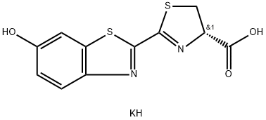 115144-35-9 结构式
基本信息
BEETLE LUCIFERIN, POTASSIUM SALT
D-(-)-2-[6'-HYDROXY-2'-BENZOTHIAZOLYL]DELTA-THIAZOLINE-4-CARBOXYLIC ACID POTASSIUM SALT
D-LUCIFERIN FIREFLY, POTASSIUM SALT
D-LUCIFERIN, MONOPOTASSIUM SALT
D-LUCIFERIN POTASSIUM SALT
D -LUCIFERIN SYNTHETIC POTASSIUM SALT
D-Luciferenpotassiumsalt
(S)-4,5-Dihydro-2-(6-hydroxy-2-benzothiazolyl)-4-thiazolecarboxylicacid,potassiumsalt
(D)-4,5-Dihydro-2-(6-hydroxy-2-benzothiazolyl)-4-thiazolecarboxylic acid, Firefly, potassium salt
D-Luciferin*K
D-LUCIFERINPOTASSIUMSALT,99%
物理化学性质
DMSO:7.5(Max Conc. mg/mL);23.55(Max Conc. mM)
Ethanol:0.25(Max Conc. mg/mL);0.79(Max Conc. mM)
PBS (pH 7.2):10.0(Max Conc. mg/mL);0.31(Max Conc. mM)
Water:32.0(Max Conc. mg/mL);100.5(Max Conc. mM)

常见问题列表
D-荧光素钾盐是荧光素酶的水溶性底物,存在于多种发光生物体中。在ATP和荧光素酶的催化作用下,D-荧光素钾被氧化,产生蓝绿色的光(560nm),当底物过量时,产生的光量子数与荧光素酶的浓度呈正相关。编码荧光素酶的Luc基因是植物、哺乳动物细胞的常用报告基因。由于没有背景干扰,因此可以很容易地检测出低至0.02 pg 水平的荧光素酶。
D-荧光素钾盐是一类在生物体中发现的能引起生物发光的杂环化合物,如萤火虫。在ATP存在下,萤光素酶将其氧化脱羧后会发光。化学研究中可用于荧光素酶的基板。
D-luciferin is the natural substrate of the enzyme luciferase (Luc), that catalyzes the production of the typical yellowgreen light of fireflies.The present review covers the synthesis of D-luciferin and derivatives or analogues that are substrates or inhibitors of the luciferase from the American firefly
Photinus pyralis
, the enzyme more frequently used in techniques of in vitro and optical imaging.
D-Luciferin exhibits a decrease in the measured K
m
in PC3M-Luc cell lysates with a K
m
of 34 μM.
Bioluminescence imaging (BLI) using the firefly luciferase (Fluc) as a reporter gene and D-luciferin as a substrate is currently the most widely employed technique. The total signal intensity is plotted against the time after D-luciferin injection to generate a time-intensity curve. In addition to the peak signal, the signals at fixed time points (5, 10, 15, and 20 min) after D-luciferin injection are determined as alternatives to the peak signal. The signal in a given time-intensity curve is normalized for the peak signal in the curve to represent the pattern of temporal changes after D-luciferin injection.
Inject with 10 μL of D-luciferin (intraperitoneally or intravenously) stock solution per gram of body weight: normally ~200 μL for a 20 g mouse for a standard 150 mg/kg injection.
Thaw D-Luciferin (either Potassium or Sodium Salt) at room temperature and dissolve in dPBS (no calcium or magnesium) to a final concentration of 15 mg/mL. Pre-wet a 0.22 μm filter by drawing through 5-10 mL of sterile H
2
O and discard water. Sterilize the D-Luciferin solution through the prepared 0.22 μm syringe filter.