TUMOR NECROSIS FACTOR
- CAS No.
- Chemical Name:
- TUMOR NECROSIS FACTOR
- Synonyms
- TSG6;α-induced protein 9;α-induced protein 6;TUMOR NECROSIS FACTOR;Anti-TSG6 antibody produced in rabbit;SixTransMembrane protein of prostate 2;Six-transmembrane epithelial antigen of prostate 4
- CBNumber:
- CB0816188
- Molecular Formula:
- Molecular Weight:
- 0
- MDL Number:
- MFCD00080185
- MOL File:
- Mol file
| storage temp. | -20°C |
|---|---|
| NCI Dictionary of Cancer Terms | tumor necrosis factor |
TUMOR NECROSIS FACTOR Chemical Properties,Uses,Production
Synthesis and release
The TNF gene transcription is induced by nuclear factor-κB (NF-κB), c-Jun, activator protein-1 (AP-1), and nuclear factor associated with activated T cells (NFAT). In the case of pathogen exposure, host immune systems detect pathogen-associated molecular patterns (PAMPs) largely through germline-encoded pattern recognition receptors (PRRs), which engage downstream of the NF-κB pathway to induce TNF mRNA. TNF mRNA contains at least three cis-elements, a 2-aminopurine response element (20 APRE), an AU-rich element (ARE), and a constitutive decay element (CDE) in the 30 untranslated region, which is required for its posttranscriptional regulation through the action of protein kinase R (PKR), RNA-binding proteins/miRNAs, and mRNA decay factors, respectively.
Receptors
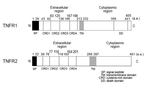
There are two types of TNF receptors: TNFR1 (also
referred to as p55, p60, TNFRSF1A, and CD120a) and TNFR2 (also known as p75, p80, TNFRSF1B, and
CD120b). The human TNFR1 gene,
TNFRSF1A, and the TNFR2 gene, TNFRSF1B, consist of
10 exons. The two receptors
are single-spanning type I transmembrane proteins and
consist of four cysteine-rich domains (CRD1 to CRD4,
from N-terminus to C-terminus), which are required for
ligand-binding, and a transmembrane region. Both
receptors form trimers, which are required for TNF binding. TNFR1 but not TNFR2 has a death domain in
its cytoplasmic region. Although sTNF binds to both
receptors with high affinity, it preferentially binds to
TNFR1 (Kd=1.9×10⁻¹¹M) compared with TNFR2
(Kd=4.2×10⁻¹⁰M). On the other hand, tmTNF
strongly activates receptor-mediated downstream signaling. The half-life of TNFR1 and TNFR2 is 180min and
10min, respectively. The primary structures of the
mouse, rat, human, cow, and pig of TNFR1 and TNFR2
are shown in Figs. 39B.S5 and 30B.S6.
Agonists and Antagonists
Purified recombinant TNF protein, selective TNFR2 antagonists (TNC-scTNFR2 and EHD2-scTNFR2). Antibodies to TNF, antibodies to TNFR1, antibodies to TNFR2, TNFR2-Fc chimeric protein, a selective human TNFR1 inhibitor based on the technology of nanobody, variable new antigen receptor (VNAR) domains against TNF, dominant-negative TNF mutants.
Clinical implications
Five anti-TNF biologics are approved for clinical use by the US Food and Drug Administration (FDA) and the European Medicines Agency (EMA) for the treatment of rheumatoid arthritis, ankylosing spondylitis, psoriasis, psoriatic arthritis, juvenile idiopathic arthritis, Crohn’s disease, and ulcerative colitis. Infliximab is a mousehuman chimera monoclonal antibody against TNF. Adalimumab and golimumab are fully humanized monoclonal antibodies against TNF. Etanercept is a soluble TNFR2-Fc chimera. Certolizumab is a PEGylated Fab fragment.
Description
The importance of TNF is highlighted by its discovery in diseases and multiple key biological processes including immune homeostasis, cell proliferation, differentiation, apoptosis, lipid metabolism, and coagulation. TNF was originally found in mouse serum after the intravenous injection of bacterial endotoxin into mice primed with a viable Mycobacterium bovis Bacillus Calmette-Guerin (BCG) strain; it was shown to cause the necrosis of tumors. Human TNF cDNA and its mRNA were identified in 4β-phorbol 12β-myristate 13α-acetate (PMA)-stimulated HL-60, a monocyte-like cell line derived from a promyelocytic leukemia. The biosynthesis of TNF was clarified by the identification of the cell surface precursor transmembrane form and its processing enzyme, TNF-α-converting enzyme (TACE).
Physical properties
Mr. 26,000 (tmTNF), 17,000 (sTNF). pI 5.8 (human sTNF). TNF is soluble in water. TNF is stable in plasma and serum for 8 h at room temperature and for 24 h at 4°C, -20°C, or -70°C.
General Description
The TNFs (Etanercept, Enbrel) are members of a family ofcytokines that are produced primarily in the innate immunesystem by activated mononuclear phagocytes. Etanercept is a dimeric fusion protein consisting of the extracellularligand-binding portion of the human 75-kDa (p75)TNF receptor (TNFR) linked to the Fc portion of human isotypeIgG1. The Fc component of etanercept contains the CH2domain, the CH3 domain, and the hinge region, but not theCH1 domain of IgG1. These regions are responsible for thebiological effects of immunoglobulins. Each etanerceptmolecule binds specifically to two TNF molecules in the synovialfluid of rheumatoid arthritis patients. It is equally efficaciousat blocking TNFα and TNFβ. The drug is indicatedfor reducing signs and symptoms and inhibiting the progressionof structural damage in patients with moderately toseverely active rheumatoid arthritis. Etanercept is also indicatedfor reducing signs and symptoms of moderately toseverely active polyarticular-course juvenile rheumatoidarthritis in patients 4 years of age and older who have had aninadequate response to one or more disease-modifying antirheumaticdrugs (DMARDS). Etanercept is also indicatedfor reducing signs and symptoms of active arthritis in patientswith psoriatic arthritis.
Clinical Use
Mutations in the TNFRSF1A1 gene are associated with autosomal dominant autoinflammatory disorder, called TNF receptor-associated periodic syndrome (TRAPS); this was first described as familial Hibernian fever (FHF). Polymorphisms in TNFRSF1B have also been identified in some patients with familial rheumatoid arthritis, Crohn’s disease, ankylosing spondylitis, ulcerative colitis, and immune-related conditions such as graft versus host disease associated with scleroderma risk. Some polymorphisms of the TNF gene promoter are observed in patients with systemic lupus erythematosus (SLE), rheumatoid arthritis, and ankylosing spondylitis.
Structure and conformation
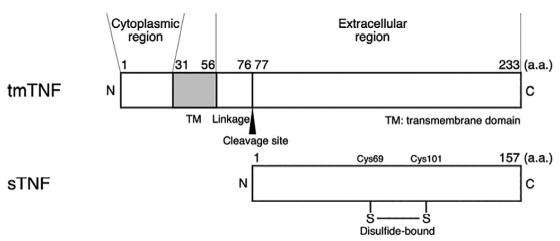
TNF exists in two forms: a type II transmembrane protein (tmTNF) and a mature soluble protein (sTNF) . The TNF protein is first produced as tmTNF on
the cell surface. tmTNF is enzymatically cleaved at its
Ala76-Val77 site by TACE and is released as sTNF. sTNF forms a noncovalently bound homotrimer that is
at least eight-fold more active than the monomer. The
TNF monomer has 10 antiparallel β-sheets and exhibits
a “jelly-roll” topology. Individual subunits form the
trimer by an edge-to-face interaction of β-sheets. sTNF
contains one disulfide bond (Cys69-Cys101), which plays
a role in its biological functions.
TUMOR NECROSIS FACTOR Preparation Products And Raw materials
Raw materials
Preparation Products
TUMOR NECROSIS FACTOR Suppliers
| Supplier | Tel | Country | ProdList | Advantage | |
|---|---|---|---|---|---|
| 3B Pharmachem (Wuhan) International Co.,Ltd. | 821-50328103-801 18930552037 | 3bsc@sina.com | China | 15848 | 69 |
| Sigma-Aldrich | 021-61415566 800-8193336 | orderCN@merckgroup.com | China | 51471 | 80 |
| United States Biological | -- | sales@advtechind.com | United States | 6106 | 58 |
| Supplier | Advantage |
|---|---|
| 3B Pharmachem (Wuhan) International Co.,Ltd. | 69 |
| Sigma-Aldrich | 80 |
| United States Biological | 58 |


